The Anatomy of the Eye
The eye is a complex structure.
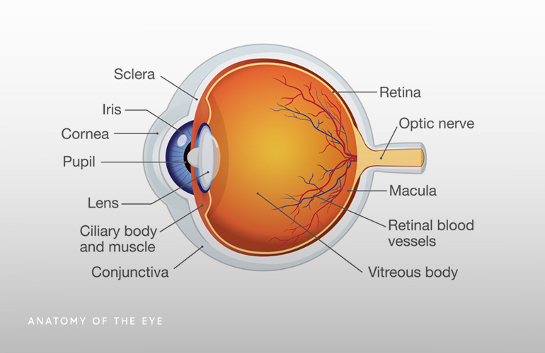
Iris
The Iris is the round colored area of the eye. Muscles located in the iris dilate or constrict the pupil to control the amount of light that enters the eye.
Lens
The lens is directly behind the pupil and sits inside the lens capsule, which suspends the lens continually changes shape to focus light onto the retina. A cloudy lens is called a cataract.
Cornea
The cornea is the clear layer of tissue that surrounds the eye. It transmits and focuses light into the eye.
Aqueous humor
The aqueous humor sits behind the cornea in a space called the anterior chamber. The aqueous humor produces fluid to maintain eye pressure and drains from the eye via a drainage angle. When the drainage becomes blocked it raises intraocular pressure (IOP) and causes glaucoma.
Retina
The retina is the part of the eye that contains light sensitive cells called photoreceptors and blood vessels that supply nutrients and oxygen to the eye structures.
Macula
The Macula is part of the retina and contains millions of photoreceptors called cones.
Photoreceptors
Photoreceptors in macula convert electrical signals from the brain to the optic nerve. Photoreceptors each have a nerve fiber that are bundled together to form the optic nerve. There are two types of photoreceptors- the cones and the rods. They change light into energy which is transmitted to the brain.
- The cones are responsible for sharp, detailed central vision and color vision and live in the macula.
- The rods perceive black and white and are responsible for night vision. Rods are located in the periphery of the retina.
Ciliary body
The ciliary body is a group of tiny muscles that make adjustments to eyesight. It changes the shape of the lens to focus near and far.
Choroid
The Choroid is a layer of blood vessels in the back of the eye that supply nutrients to the eye.
Sclera
The sclera is the white part of the eye. It is a strong tissue that covers the surface of the eyeball. There are six muscles that are attached to the sclera and control eye movement. Th sclera is also covered with tissue called the conjunctiva which is a clear membrane.
Lacrimal glands
The lacrimal glands sit inside the eye orbit and produce the watery part of tears. Tears keep the eye lubricated.
Meibomian glands
The meibomian glands sit inside the lower eye lashes and produce oil as part of the tears, to prevent the watery tears from drying quickly.
Optic nerve
The Optic nerve is the neve that carries electrical signals from the eye to the brain and from the brain to the eye.
Retina
The retina is light sensing layer of nerves lining the back of the inside of the eyeball. We see when light strikes the retina and creates electrical impulses which are sent through the optic nerve to the brain and interpreted as eye sight.
Macula
The macula is the part of the retina that contains the light sensitive cells that provide the ability to see fine details in the center of the visual field. Peripheral vision occurs in the peripheral parts of the retina. Macular degeneration is a disease caused by deterioration of the macula.
Fovea
The fovea is in the center of the macula and provides for sharp vision.
Vitreous humor
The vitreous humor is a clear jelly like substance that fills the cavity of the eyeball delivers nourishment to the eye and helps the eye to keep its shape.

 / 53 Reviews